Rectal Prolapse Surgery
Conference Name: Webinar on Piles Surgery
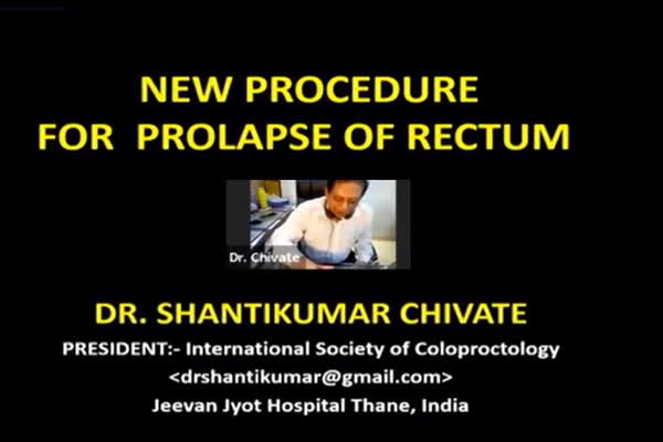
Conducted by: Dr. Shantikumar Chivate
Surgeon(s)/Speaker(s): Dr. Shantikumar Chivate
Surgical Procedure: Colorectal-Rectopexy for recurrent prolapse
Location: Meditorch's Virtual Meet
Indexsteps
1. Patient position : The patient is placed in a steep Trendelenburg position under regional anesthesia.
2. Marking : First, the rectal prolapse should be confirmed with the apex marked with a suture.
3. Prolapse reduction : Prolapse reduction and irrigation of the rectum is done using normal saline.
4. metal proctoscope introduced : Next, the side view, wide-operating metal proctoscope is introduced in the anal cavity such that it is placed adjacent against the sacrum.
5. prolapsed rectum repositioned : The prolapsed rectum should be repositioned so that the operation could be performed through the side window of the proctoscope.
6. Suture inserted : A No. 01 PDS suture (40-mm round body needle) is inserted through the upper right-hand edge of the window across the rectum.
7. First needle placement : The first needle is to be placed as high as possible against the sacrum at a minimum lateral distance of 3 cm from the midline, deemed the upper margin of a triangle of vascular safety just below the sacroiliac joint.
8. Needle brought to an identical position : Once this needle is correctly inserted it is brought to an identical position on the other side of the sacrum by passing in direct contact with the bone in order to avoid a presacral venous injury.
9. Needle turned back : The needle is then turned back and grasped through the left edge of the rectal wall so that 2/3 of the circumference of the rectal wall is incorporated into the presacral fascia.
10. Poition and security of the stitch should to be confirmed : The position and security of the stitch should be confirmed by pulling the suture downwards and ensuring that the rectum remained fixated at that point.
11. Rectal fixation : Rectal fixation is to be performed from approximately the 3rd sacral vertebra to the sacrococcygeal joint with typically 4 to 5 sutures being required.
12. Suturing : Each suture should be tied using a laparoscopy knot pusher.
13. The lax part of the posterior rectal wall is elevated : The lax part of the posterior rectal wall is elevated with Babcock forceps and a 20 cm long, 20-gauge needle must be inserted through the rectal wall into the presacral space with the position of the needle confirmed with transrectal ultrasound.
14. presacral sclerosant injection : The 2-mL solution of sodium tetradecyl sulfate/polidocanol (30 mg/mL) is mixed with 8 mL of air and shaken into a foam for injection (presacral sclerosant injection) under endosonographic guidance.
port positions
1. Steep Trendelenburg position : Steep Trendelenburg position
pre_post_measures
1. Preoperative investigations : *A detailed clinical history and examination for confirmation of FTRP *Patient’s age, symptoms, medical history, physical examination findings, imaging studies and laboratory test results *Recording of the presence of symptoms of bleeding, prolapse, pain, and burning *Assessment according to the Agachan constipation score and the Pescatori incontinence score
2. Postoperative measures : ▪ The patient should be continued on antibiotics for 5 postoperative days and be provided oral analgesia as required. ▪ The patients must be kept in the bed in a slight Trendelenburg position with a postoperative urinary catheter for 4 days and managed with low dose heparin and anti-embolic stockings. ▪ A liquid diet is advised until the 5th postoperative day post which conversion to a normal diet can be done with the use of lactulose as required. ▪ On discharge, patients should be advised against excessive straining at stool and encouraged to drink plenty of fluids, to eat a diet with a high leafy green vegetable content and where possible to practice Yoga and mindfulness strategies
3. Preoperative measures : ▪ Patient should be given clear liquids and 3 doses of osmotic cathartic lactitol at 4 hourly intervals on the prior day of the procedure. ▪ Metronidazole, ceftriaxone, and sulbactam should be administered intravenously on induction.
Surgical Instruments
1. Dr. Chivate's Proctoscope
2. No. 01 PDS suture
3. 40-mm round body needle
4. Knot Pushers
5. 5mm Needle Holder
6. Babcock forceps
7. 20 cm long 20-gauge needle
8. polidocanol (30 mg/mL
9. Wet gauze soaked in saline solution
